Nuclear Medicine Physics for FRCR Part 1: Gamma Cameras Explained
Nuclear medicine physics for FRCR Part 1 explained, with gamma cameras, collimators, resolution, sensitivity, and the exam points candidates miss most.
Answer First
Nuclear medicine physics in FRCR Part 1 is mainly about how gamma cameras form images, how collimators trade sensitivity for resolution, and why the patient is the radiation source.
Key Facts
- A gamma camera detects photons emitted from a radiotracer inside the patient.
- Collimators improve directional accuracy but reduce sensitivity.
- Resolution versus sensitivity is one of the most frequently tested nuclear medicine trade-offs.
- Radiation safety in nuclear medicine differs from X-ray-based imaging because the source is inside the patient.
Practice
Apply this with real FRCR Part 1 questions
Reinforce today’s topic with targeted practice in the Spotters Academy app.
Nuclear medicine physics is one of the most intimidating sections of FRCR Part 1 physics - especially topics like gamma cameras, collimators, and resolution vs sensitivity.
FRCR candidates often struggle with nuclear medicine physics not because it is conceptually difficult, but because it is often taught as fragmented facts rather than as a logical imaging system.
This guide explains nuclear medicine physics for FRCR Part 1, with a clear focus on gamma cameras, what examiners actually test, and how to study this topic efficiently without over-memorising.
This guide is aligned with the Royal College of Radiologists FRCR Part 1 physics syllabus.
Why Nuclear Medicine Physics Matters for FRCR Part 1
Nuclear medicine physics is tested because it assesses:
-
understanding of image formation
-
radiation detection principles
-
resolution vs sensitivity trade-offs
-
patient and staff radiation safety
Questions are usually concept-based, not calculation-heavy.
What Does FRCR Expect You to Know in Nuclear Medicine Physics?
For FRCR Part 1, you are expected to understand:
-
basic gamma camera components
-
how images are formed
-
factors affecting image quality
-
common misconceptions and traps
You are not expected to memorise engineering-level detail.
Gamma Cameras Explained for FRCR (Step by Step)
1️⃣ What Is a Gamma Camera?
A gamma camera detects gamma photons emitted from radiotracers inside the patient and converts them into an image representing tracer distribution.
Unlike X-ray or CT imaging:
-
the radiation source is inside the patient
-
photons are detected, not transmitted
This is a fundamental conceptual difference often tested in exams.
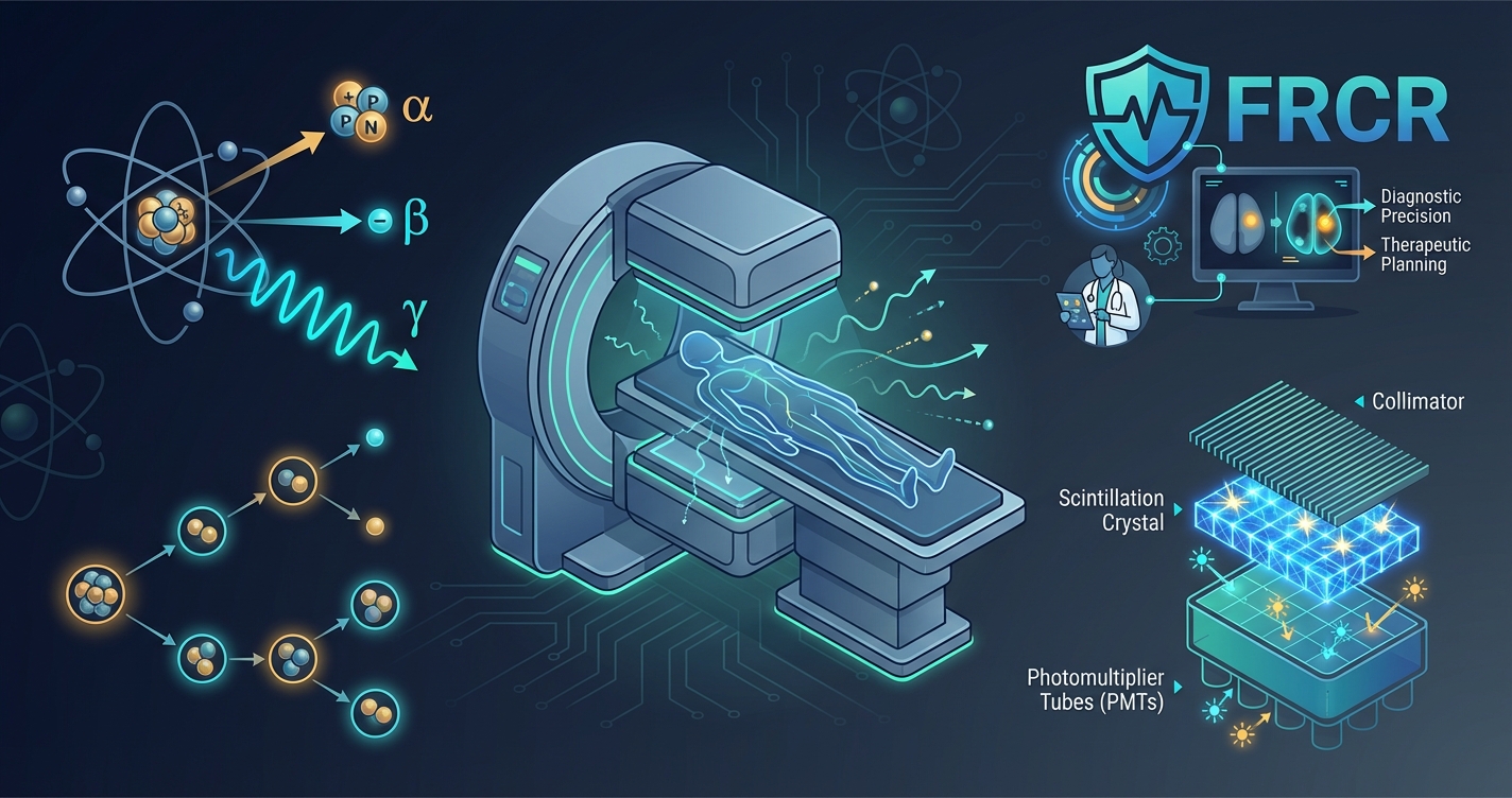
2️⃣ Key Components of a Gamma Camera
Collimator
-
Determines direction of incoming photons
-
Rejects scattered photons
-
Trades sensitivity for spatial resolution
FRCR pearl:
Better resolution = lower sensitivity (and vice versa).
Scintillation Crystal
-
Converts gamma photons into light
-
Usually sodium iodide (NaI)
-
Thickness affects sensitivity and resolution
Photomultiplier Tubes (PMTs)
-
Convert light into electrical signals
-
Allow localisation of photon interaction
Resolution vs Sensitivity: A High-Yield FRCR Topic
This relationship is frequently tested.
-
High resolution collimator
-
Better spatial resolution
-
Lower sensitivity
-
Longer imaging time
-
-
High sensitivity collimator
-
More counts
-
Poorer resolution
-
Faster imaging
-
FRCR questions often test trade-offs, not definitions.
Common Nuclear Medicine Physics Pitfalls in FRCR
Common FRCR nuclear medicine mistakes include:
-
confusing gamma cameras with PET detectors
-
assuming higher energy always improves resolution
-
forgetting the role of the collimator
-
over-memorising crystal physics
Most errors come from misunderstanding image formation.
Radiation Safety in Nuclear Medicine (FRCR Focus)
Key principles:
-
Radiation source is the patient
-
Distance and time are critical
-
Shielding differs from X-ray-based modalities
These concepts are often tested indirectly and overlap with our guide to radiation dosimetry for FRCR Part 1.
Nuclear Medicine Physics for FRCR: At a Glance
| Topic | Exam Priority |
|---|---|
| Gamma camera principle | Very high |
| Collimators | Very high |
| Resolution vs sensitivity | Very high |
| Crystal & PMTs | Moderate |
| Radiation safety | High |
How to Study Nuclear Medicine Physics for FRCR Part 1
-
Focus on conceptual flow
-
Use diagrams to visualise photon paths
-
Practice True/False style questions
-
Revise trade-offs repeatedly
You do not need to master PET or advanced reconstruction algorithms for Part 1.
Frequently Asked Questions (FAQ)
Is nuclear medicine physics heavily tested in FRCR Part 1?
It appears regularly but is usually concept-based rather than calculation-heavy.
Do I need to memorise gamma camera components?
You need to understand their function, not memorise technical specifications.
Are collimators important for the exam?
Yes. Very important.
Is PET physics required at the same depth?
No. PET is usually tested at a more superficial level.
What is the most common mistake candidates make?
Not understanding that the patient is the radiation source.
Final Takeaway
Nuclear medicine physics becomes manageable when you:
-
understand image formation
-
focus on trade-offs
-
avoid unnecessary detail
For FRCR Part 1, clarity beats memorisation. To see how nuclear medicine fits with the other physics topics and plan your revision, follow the FRCR Part 1 physics study guide.
Author
Dr B Gayathri Priyadharshinee
FRCR Radiologist & Educator
Dr Gayathri mentors radiology trainees for international exams, focusing on physics clarity, exam logic, and high-yield preparation strategies.
For a structured topic-by-topic plan, see our FRCR Part 1 physics revision guide.
Sources and further reading
Checked on 10 June 2026.
Tags
Sources
Dr. Gayathri Priyadharshinee
Expert content from the Spotters Academy team. We're dedicated to helping radiologists succeed in their FRCR Part 1 examination.
Ready to ace your FRCR Part 1?
Join thousands of successful candidates who prepared with Spotters Academy.
Start Free Trial